Comprehensive Guide to Urinary Tract Imaging
Importance of Accurate Urinary Tract Imaging
Accurate imaging of the urinary tract helps diagnose and treat kidney and urinary disorders effectively. Many people think x-rays are the primary imaging tool, but they work best for detecting certain types of kidney stones and monitoring their size and position. However, not all kidney stones are visible on x-rays, so other imaging methods are often more reliable and widely used.
Diagnostic Tests for Kidney and Urinary Tract Disorders
To diagnose and manage kidney and urinary tract conditions, various diagnostic tests are available. Below is an overview of the most common imaging methods, their advantages, and their specific applications.
X-Ray Imaging
Many believe x-rays are ideal for urinary tract problems, but this is not entirely accurate. X-rays are useful for detecting some kidney stones and tracking their movement. However, several types of stones, such as uric acid stones, are invisible on plain x-rays. Doctors often combine x-rays with other advanced imaging techniques for a complete diagnosis.

Ultrasound Imaging
Ultrasound is a popular diagnostic tool for urinary tract issues because it is safe, cost-effective, and widely available. Key benefits include:
- Avoiding harmful ionizing radiation.
- Offering real-time imaging, allowing on-the-spot evaluation.
- Effectively detecting stones, swelling, or blockages.

Technicians can also use Doppler ultrasound to assess blood flow, aiding in the diagnosis of testicular conditions, erectile dysfunction, and kidney issues.
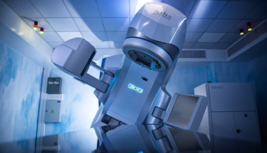
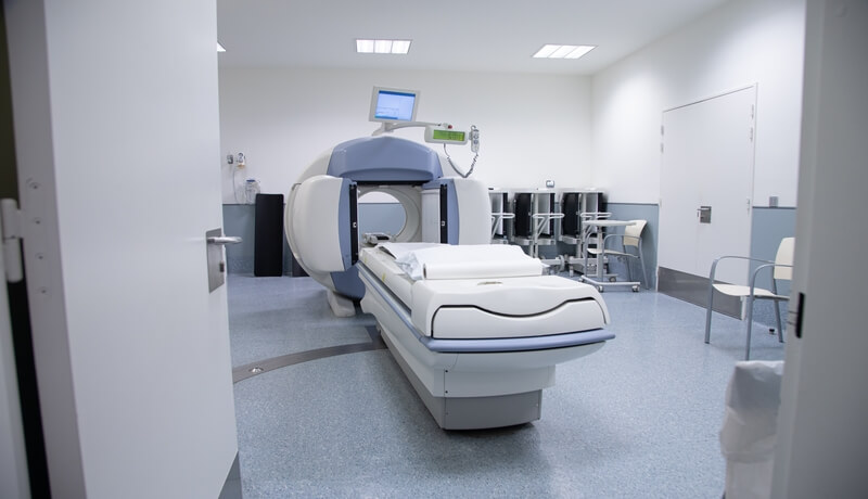
Computed Tomography (CT)
CT scans provide detailed images of the urinary tract and surrounding areas. They are particularly useful for detecting kidney stones, tumors, and structural abnormalities. CT angiography, a less invasive alternative to standard angiography, allows doctors to assess vascular conditions. However, CT scans involve high radiation exposure and may require contrast agents, which could harm the kidneys or cause allergic reactions.
Magnetic Resonance Imaging (MRI)
MRI offers clear images of the urinary tract and nearby tissues without exposing patients to radiation. It excels at evaluating blood vessels and soft tissue conditions that require detailed imaging. However, MRI is not effective for detecting urinary stones. Additionally, the contrast agents used in MRI are unsuitable for individuals with weak kidney function, as they can lead to serious complications.

Intravenous Urography (IVU)
Intravenous urography (IVU), also called intravenous pyelography (IVP), involves injecting a contrast agent into a vein to highlight the kidneys, ureters, and bladder on x-rays. Though largely replaced by CT urography, IVU remains useful in specific cases, such as identifying recurrent infections or structural abnormalities.
Retrograde Urography
This imaging technique directly injects a contrast agent into the urinary tract through the bladder. Doctors use retrograde urography to identify scarring, tumors, or blockages in the urinary system. It is especially helpful for patients with poor kidney function who cannot tolerate intravenous contrast agents.
Percutaneous Antegrade Urography
Doctors use this method to inject a contrast agent directly into the kidney through a small incision in the back. It is performed when retrograde urography is not feasible or when a nephrostomy tube is already in place to treat blockages caused by stones or tumors.
Cystography and Cystourethrography
In cystography, a contrast agent is injected into the bladder to detect structural abnormalities or injuries. Cystourethrography, on the other hand, examines both the bladder and urethra to identify blockages or strictures. A variation called voiding cystourethrography evaluates urine flow and reveals any narrowing in the urethra.
Retrograde Urethrography
Retrograde urethrography involves injecting a contrast agent directly into the urethra to diagnose injuries or narrowing. It is commonly used in cases of trauma to ensure safe catheterization or further treatment.
Positron Emission Tomography (PET)
PET imaging uses specialized agents to detect prostate cancer and other tumors in the genitourinary system. This advanced technique is particularly effective for identifying metastases and assessing kidney or testicular cancers.
Radionuclide Scanning
Radionuclide scanning uses a gamma camera to track radiation from a radioactive tracer injected into the body. Doctors use this test to evaluate kidney blood flow and assess how well the kidneys filter and excrete urine.
Angiography
Angiography involves injecting a contrast agent into blood vessels to detect abnormalities, such as fistulas or blockages. This procedure is crucial for diagnosing and treating vascular conditions affecting the urinary tract. However, complications like bleeding or adverse reactions to the contrast agent can occur.

Improving Patient Outcomes with Advanced Imaging
Using advanced urinary tract imaging methods, healthcare professionals can accurately diagnose and treat kidney and urinary disorders. By choosing the right test for each condition, doctors enhance patient care and overall outcomes.
Frequently Asked Questions
FAQs with Answers
-
What are the risks of using contrast agents in urinary tract imaging?
Contrast agents can occasionally cause allergic reactions, ranging from mild itching to severe anaphylactic responses. In some cases, they may lead to kidney damage, especially in patients with pre-existing kidney issues or diabetes. To minimize risks, doctors assess the patient’s medical history and kidney function before administering contrast agents. Alternatives like ultrasound or MRI (without contrast) may be recommended for high-risk patients. -
How does Doppler ultrasound improve the detection of urinary tract disorders?
Doppler ultrasound evaluates blood flow in real-time, which is particularly helpful for diagnosing conditions like renal artery stenosis or testicular torsion. By analyzing the movement of blood through vessels, it provides additional insights that standard ultrasound cannot, such as identifying blockages or abnormal blood circulation in the urinary tract. -
When should retrograde urography be chosen over intravenous urography?
Retrograde urography is preferred when intravenous contrast agents are contraindicated due to poor kidney function. It is also ideal for detecting localized abnormalities, such as scarring, strictures, or tumors, where a more targeted approach is needed. This method is often used during urologic procedures to ensure precise imaging of the ureters and renal pelvis.
For further assistance, visit our dedicated consultation page: Contact HOMED for Consultations.